Sponsor Area
Make a comparison and write down ways in which plant cells are different from animal cells?
Difference between the plant cell and the animal cell are:
|
Plant Cell |
Animal Cell |
|
1. The outermost covering of the plant cell is the cell wall which is formed of cellulose. 2. Plastids (e.g., chloroplast) present. 3. Large vacuole present. 4. Centrioles are absent but polar caps are present. 5. Golgi apparatus is in the form of sub-units, called dictyosomes. |
1. The outermost covering is the plasma membrane. 2. Plastids absent. 3. Small or no vacoules are present. 4. Centrioles are present within centrosome. 5. Prominent and highly complex Golgi apparatus is present near nucleus. |
How is a prokaryotic cell different from a eukaryotic cell?
|
Prokaryotic Cell |
Eukaryotic Cell |
|
1. Cell size is generally small (1-10 μm) 2. Nucleus is not bounded by a membrane and it is called nucleoid. 3. Only a single chromosome is present. 4. Nucleolus is absent. 5. Membrane bound cell organelles are absent. 6. Cell division by fission or budding (no mitosis). |
1. Cell is generally large (5-100 (μm) 2. Nuclear material is surrounded by a nuclear membrane. 3. More than one chromosome are present. 4. Nucleolus is present. 5. Cell organelles bounded by membrane are present. 6. Cell division mitotic or meiotic. |
What would happen if the plasma membrane ruptures or breaks down?
The plasma membrane provides shape and support to the cell. It is a selectively permeable membrane and keeps a check on the movement of substances in and out of the cell by the process of diffusion or osmosis. Thus, if the plasma membrane is ruptured it cell contents will be in direct contact with the enviroment and movement of substances will no longer be selective. Free movement may lead to the death of the cell.
What would happen to the life of a cell if there was no Golgi apparatus?
Golgi apparatus perform the function of storage, packaging and dispatch of substances synthesised by endoplasmic reticulum. It transports the material in and out of the cell. It is also reponsible for the formation of lysosomes. Therefore, if there were no golgi bodies then there would be no storage, packaging and transport of material. The formation of lysosomes will not take place in the absence of Golgi apparatus. All this will lead ultimately to the death of Golgi apparatus.
Which organelle is known as the powerhouse of the cell? Why?
Mitochondria are known as ‘Power House’ of the cell.They are called so because they produce energy in the form of ATP molecules. ATP is the energy currency of the cell. It is stored and used for making new compounds and mechanical work.
Where do the lipids and proteins constituting the cell membrane get synthesized?
The proteins are synthesised on the Rough Endoplasmic Reticulum (RER) and the ribosomes attached to it. While the lipids are synthesised on the Smooth Endoplasmic Reticulum (SER).
How does an Amoeba obtain its food?
What is osmosis?
Osmosis is the passage of water from a region of its high concentration to the region of its lower concentration through a semipermeable membrane.
Carry out the following osmosis experiment:
Take four peeled potato halves and hollow each one out to make potato cups. One of these potato cups should be made from a boiled potato. Put each potato cup in a trough containing water. Now,
(a) keep cup A empty
(b) put one teaspoon sugar in cup B
(c) put one teaspoon salt in cup C
(d) put one teaspoon sugar in boiled potato cup D.
Keep this setup for two hours. Then observe the four potato cups and answer the following :
(i) Explain why water gathers in the hollowed portion of B and C?
(ii) Why is potato A necessary for this experiment?
(iii) Explain why water does not gather in the hollowed out portions of A and D.
a) Water gathers in the hollowed portion of potato B and C because the plasma membrane of potato cup act as semipermeable membrane and the concentration gradient causes the water to move from the cell to the hllowed portion by the process of osmosis.
(b) Potato cup A is necessary as it acts as a control. This shows that osmosis occurs only when there is concentration gradient and a living semi permeable membrane.
(c) (i) In the potato cup A, there is no concentration gradiesnt for osmosis to occur.
(ii) In the potato cup D, the semi permeability of potato membrane is lost due to boiling. So, no water movement occurs.
By which instrument you can see internal structure of onion peel cell?
Compound microscope.
Do different organisms have different types of basic building units ? Briefly explain.
No, all organisms have basic building units called cells. Cells are the basic structural and functional unit of any living cell.
Can you find out more names of some of the unicellular organisms and fungi, other than given in your book?
Plasmodium (Malaria parasite) and Yeast are unicellular organisms.
Sponsor Area
Name three features which are found almost in every cell.
The three features found in almost every cell are plasma membrane, nucleus and cytoplasm.
What is the function of cell wall and plasma membrane?
Cell wall provides shape and protection to plant cell.
Cell membrane provides protection, shape and flexibility to the cell. It acts as a selectively permeabl Allows only selected materials to move in and out of the cell through the process of diffusion or osmosis.
What are cell organelles?
The memberane bound little structures found within the cell are called as cell organelles. They are the functional units of a cell.
Define diffusion.
Diffusion is the spontaneous movement of a substance from a region of its high concentration to the region where its concentration is low. For example the movement of CO2 or O2 across the cell membrane.
Why is plasma membrane called a selectively permeable membrane?
Plasma membrane regulates the entry and exit of materials in a cell. It allows the movement of only certain substances while prevents the others from doing so. Thus, it is called selectively permeable membrane.
Define osmosis.
The movement of water from its region of higher concentration to the region of its lower concentration through a semipermeable membrane is called osmosis.
What is endocytosis? Give one example.
Endocytosis is the process of engulfing food and other materials by a cell from the external environment. The flexibility of the cell membrane helps in the process For example - Amoeba engulf food through this process.
What will happen if an animal or plant cell is put into a solution of sugar or salt prepared in water?
Depending on the concentration difference between the cell and the solution the following three things could happen:
(i) If the solution surrounding the cell is less concentrated (hypotonic) than the cytoplasm, the water will move into the cell, i.e., the cell will gain water and swell.
(ii) If the solution has the same concentration as that of cytoplasm of cell, there will be no net movement of water across the cell membrane, i.e., no gain or loss of water from the cell.
(iii) If the solution is highly concentrated (hypertonic) as compared to the cytoplasm, i.e., the solution is concentrated, the cell will lose water by osmosis.
What will happen when:
(i) an egg without shell placed in concentrated salt solution for 5 minutes.
(ii) an egg without shell placed in pure/distilled water for 5 minutes. Give reason in brief.
The following results are observed
(i) When an egg without shell is placed in concentrated salt solution for 5 minutes - The egg shrinks due to loss of water by the process of osmosis.
Reason. The egg membrane acts as semipermeable membrane. The water concentration inside the cell is more therefore water moves out of the cell.
(ii) When an egg without shell placed in concentrated salt solution for 5 minutes. - The egg gain water and swells.
Reason. The concentration of water is much more on the outside thus water moves into the egg cells through egg cell membrane by osmosis.
What is chromatin material?
Chromatin material is the entagled mass of thread like structures which are organised to formchromosomes during the time of cell division.
What is cytoplasm?
Cytoplasm is the fluid content present inside the cell. It houses all the cell organelles.
Sponsor Area
Name the scientists who formed primitive microscope in the 17th century respectively.
Robert Hooke
What is the location and main component of cell wall?
Cell wall is a rigid structure found outside the plasma membrane of the plant cells. It is composed of cellulose - a fibrous polysaccharide.
What is the significance of cell wall in plant cell?
Cell wall performs the following functions in the plant cell:
(i) It gives a definite shape to the cell.
(ii) It provides rigidity and strength to the cell.
(iii) It protects the cell.
(iv) It also withstands the osmotic pressure which is developed by cell contents and protects the cell from bursting.
Distinguish between plasma membrane and cell wall.
|
Plasma Membrane |
Cell Wall |
|
1. It is living. 3. It is found in both plant and animal cells. 4. It is semi-permeable. 5. It is soft and elastic. |
1. It is non-living. 3. It is found in plant cells only. 4. It is permeable. 5. It is hard and rigid. |
Explain the structure of nucleus?
Nucleus. It is the control centre of the cell. It is covered by an double layered envelope called the nuclear membrane . There are numerous pores in the nuclear membrane for the transfer of materials. It consists of a dense region called nucleolus. The nucleus has chromatin which forms the chromosomes during cell division. Chromosomes consists of DNA and protein. The genes are functional unit of DNA which are responsible for the hereditary characters of an organism. 
What are genes? Where are they found?
Genes are the functional segments of DNA (deoxyribonucleic acid) molecule. Genes are arranged in a single linear order along the chromosome. They are formed due to condensation of chromatin material at the time of cell division. It is the unit of heredity which is transferred from a parent to offspring. Genes are found inside the nucleus.
What do you observe when you see temporary slide of surface of human cheek material under microscope ? How does the shape of the cells look like? Draw labelled diagram of it.
When we observe the cheek cells under microscope we observe that the cells are flat and polygonal in shape with irregular boundaries. They are arranged edge to edge forming a delicate sheet of cells. There is a darkly stained dot like structure which represents the nucleus.

What are chromosomes?
Chromosomes are rod shaped structures found in the nucleus of the dividing cell. Chromosome is composed of DNA and protein. It contains information for inheritance of features from parents to next generation.
Draw labelled diagram of a onion peel cell. What type of cell is it? Give reason in support of your answer.

Onion peel cell is an eukaryotic cell.
Reason. Onion peel cell contains a well defined nucleus enclosed within a nuclear membrane.
What is a eukaryotic cell?
A eukaryotic cell has a definite nucleus that is a nuclear membrane bound nucleus. It also contains membrane bound organelles in its cytoplasm.
What is nucleoid?
Nucleoid is the undefined nuclear region containing only nucleic acids, in the prokaryotic cells. It is not bounded by a nuclear membrane and lies in direct contact with the cytoplasm.
Give two examples of prokaryotic organisms.
Bacteria and Cyanobacteria (earlier called blue green algae) are prokaryotic unicellular organisms.
What is a prokaryotic cell?
A cell which does not have a definite nucleus that is the nucleus is not bounded by a nuclear membrane is called as a prokaryotic cell. It has an undefined nuclear region called nucleoid.
Write three main points of cell theory as expressed by Schleidn, Schwan and Virchow.
Cell theory was proposed by Schleidn and Schwann and Rudolf virchow. It states that:
(i) the cell is the basic unit of life.
(ii) all the plants and animals are composed of cells and that
(iii) all cells arise from pre-existing cells.
List four major functions of a cell.
Functions of Cell:
(i) It acts as the funtional and structural unit.
(ii) Synthesis and transport of substances.
(iii) Energy Production.
(iv) Cellular replication.
What is the function of an endoplasmic reticulum?
Endoplasmic reticulum forms a network system and helps in the transport of substances in and out of the cell. It also provides the attachment sites for ribosomes which are the site for protein synthesis. The endoplasmic reticulum helps in the synthesis of lipids and fats.
Why do viruses do not show characteristics of life?
Viruses lack any membranes. Hence, they do not show characteristics of life until they enter a living cell and use its cell machinery to multiply.
Write one function of each—
(i) rough endoplasmic reticulum and
(ii) smooth endoplasmic reticulum.
(i) Rough endoplasmic reticulum (RER) has ribosomes on its surface and therefore helps in the synthesizing proteins.
(ii) Smooth endoplasmic reticulum (SER) helps in the manufacture of fat molecules or lipids.
What is endoplasmic reticulum? Write its main functions.
Endoplasmic Reticulum is a membranous network, enclosing a fluid-filled lumen. Its main functions are:
(i) Synthesis of proteins (Rough ER)
(ii) Synthesis of lipids and their secretion (SER).
(iii) Secretion of enzymes and hormones inside the cell or when secreted out of the cell.
(iv) ER also produces substance for new cellular parts (especially cell membrane).
(v) ER provides internal support (mechanical support) to the cell.
Briefly explain the structure of Golgi apparatus.
Golgi apparatus consists of flattened membranous sac like structures called Cisternae. These are placed one above the other (stacked) in parallel rows. These membranes are often in connections with membranes of ER.
What are the functions of Golgi apparatus?
These are functions of Golgi apparatus:
(i) Golgi apparatus is the secretory organelle of the cell. It packages and dispatches the material synthesized in the cell to intracellular (plasma membrane and lysosomes) and extracellular targets.
(ii) Golgi complex is also involved in the formation of lysosomes and peroxisomes.
(iii) Golgi apparatus is also involved in the synthesis of complex substances such as polysaccharides, glycoproteins etc from simple substances.
(iv) It helps in storage, modification and packaging of synthesised substances in the vesicles.
What are lysosomes?
Lysosomes are membrane bound vesicles act as waste disposal system of the cell. Lysosymes have digestive enzymes which help in digesting foreign material as well as worn-out cell organelles. The enzymes breakdown the organic substances. Lysosymes are called the suicide bags of the cell as they help in the digestion of damaged, worn out parts of its own cell.
What are the functions of lysosomes?
Functions of Lysosomes:
(i) Extracellular digestion. Sometimes lysosome enzymes are released outside the cell to break down extracellular material.
(ii) Digestion of foreign material. Lysosomes also destroy any foreign material such as bacteria.
(iii) Cellular digestion. Lysosomes help in the breakdown and digestion of various substances inside the cell
(iv) Waste disposal - Lysomes acts as suicidal bags and digest any damaged or worn out cells of the cell they are present in.
What is the structure of mitochondria?
Mitochondria are rod-shaped cell organelle found in the cytoplasm. Each mitochondrion is a double membrane bounded structure. The outer membrane of mitochondrion is smooth. But the inner membrane of the mitochondrion is folded inwardly into the matrix of mitochondrion forming finger-like projections called Cristae. Cristae greatly increase the surface area of the inner membrane. Mitochondria also has its own DNA. It helps in the synthesis of ATP which is the energy currency of the cell.

What are functions of Mitochondria?
Functions of Mitochondria are:
(i) They help in cellular respiration.
(ii) The stepwise released energy by the oxidation of food (usually glucose) is converted into chemical energy in the form of ATP (adenosine triphosphate) molecules.
(iii) They have their own DNA and proteins hence they prepare their own proteins.
Expand the term ATP. What is use of ATP?
ATP stands for Adenosine triphosphate.
ATP is the energy currency of the cell. The stored ATP is used for the synthesis of new chemical compounds and mechanical works.
What are plastids?
Plastids are found in plant cells only. Plastids are double membrane bounded structures. It has an extensive and numerous membrane layers embedded in a material calle stroma. They also have their own DNA and ribosomes. . TThey are of two types of plastids - chromoplasts and leucoplasts.
What are the leucoplasts and their functions?
Leucoplasts are colourless or white plastids and do not contain any pigment. Function. Leucoplasts are involved in formation and storage of starch and oil drops. For example, Amyloplasts found in potato tuber store starch.
What are chromoplasts? Which one is the most important and why?
Chromoplasts are coloured plastids i.e., They may contain pigment of different colours. For example, Blue green chromoplasts found in blue green algae.
The most important chromoplasts are chloroplasts which contain green pigment, known as chlorophyll. Chloroplasts are the kitchen of the cell and are very important because they trap solar energy and prepare food for all non-green organisms, directly or indirectly. The process of preparing food by green plants is known as Photosynthesis.
Other non-green chromoplasts are responsible for characteristic colour of flowers and fruits.
What is plasmolysis?
The shrinkage or contraction of the protoplasm away from the cell wall when kept in an hypertonic solution is called plamolysis.
What are vacuoles?
Vacuoles are are single membraned storage sacs for solid or liquid content. The membrane covering the vacuoles is called tonoplast. The fluid in the vacuoles is called cell sap. In most plant cells, vacuole is big and centrally placed. The liquid cell sap of vacuole provide turgidity and rigidity to plant cells. In animal cells either they are absent or are small.
Sponsor Area
What is the difference between a unicellular and multicellular organism ? How is multicellularity advantageous over unicellularity?
In an unicellular organism have a single cell and all functions are performed by the same cell. Multicellular organisms have more than one cells. The cells are present in colonies or are differentiated and specialsed into groups that carry out specific functions. Multicellularity provides division of labour. This helps the organisms to perform various life processes in a better way.
Draw labelled diagrams of animal and plant cells to show their internal structure
Internal structure of Plant and Animal cell.

Write one important function each of mitochondria and ribosomes.
|
Organelle |
Function |
|
(i) Mitochondria |
(i) Site of cellular respiration and ATP formation |
|
(ii) Ribosomes |
(ii) Site of protein synthesis. |
Lysosomes are often called ‘suicide bags Why?
Lysosomes contain digestive or hydrolytic enzymes. When a cell gets damaged or worn out the lysomes burst releasing the enzyme that digest their own cell. Thus they are called as ‘suicide bags’.
Name some organelle which are met with only in animal cells and those which occur only in plant cells.
(i) Structure found only in animal cells:centrosomes, golgi apparatus.
(ii) Structures found only in plant cells : cell wall, plastids, polar caps, dictyosomes and big vacuoles.
Name the cell organelle which are known as:
|
(i) Control centre |
(ii) |
Demolition squads |
|
(iii) Export firms |
(iv) |
Power house of the cell |
|
(v) Kitchen of the cell |
(vi) |
Internal transport sys |
Write a note on the structure of cell?
Cell is the fundamental unit of a living organism.
The cell is enclosed by an outer Plasma membrane which is selectively permeable. Plant cells have an additional cell wall located outside the plasma membrane. The cell is filled with a translucent viscous substance called the cytoplasm in which are embedded the organelles. The control centre of the cell is the nucleus, it contains all the information necessary for the cell to function and to reproduce further cells of the next generation.
Surrounding the nucleus is the Endoplasmic reticulum (ER) which may have the ribosomes which are the site of protein synthesis. Ribosomes, a granular structures, also found in cytoplasm.
The power house of the cell is the mitochondria which helps in releasing energy by the oxidation of food in cell.
There are flat membranous secretory structures in the cell the Golgi bodies. In plant cells there is an additional structure the chloroplast which is the site for photosynthesis.
Cells also contain lysosomes which are also called suicide bags. They digest and remove the unwanted debris of the cell. Centriole helps in cell division. Cytoplasm also contains vacuoles filled with cell sap.
In plant cells vacuole are present which act as storage sacs. Vacuoles are large in plant cells nd small or non existeant in animal cells.
What are the three functional regions of cell?
The main regions of all cells are:
(i) The plasma membrane
(ii) The nucleus and
(iii) The cytoplasm.
Can you name the two organelles that you have studied about, which contain their own genetic material?
The two cell organelles that contain their own genetic material are:
(i) Mitochondria and (ii) Chloroplast.
If the organization of a cell is destroyed due to some physical or chemical influence, what will happen?
Each cell has got cell organelles and each cell organelle performs a special function, e.g., making of new material, removal waste from the cell, release of energy etc. If the organization of a cell is destroyed, the functioning of the cell organelle will be disturbed, and cell will not be able to perform the basic functions. Thus, cell will ultimately die.
Define the term ‘tissue’.
Tissue is the group of cells similar in structure that work together to achieve a particular function. For example - blood, bone , xylem and phloem etc
How many types of elements together make xylem tissue? Name them.
Xylem formed of four types of elements. They are (i) tracheids (ii) vessels (iii) xylem parenchyma (iv) xylem phloem.
How are simple tissues different from complex tissues?
Simple tissues are made of one type of cells which coordinate to perform a common function. For example parenchyma, epidermis etc
Complex tissues are made of more than one type of cells. All these coordinate to perform a common function. For example Vascular tissues.
Differentiate between Parenchyma, Collenchyma and Sclerenchyma on the basis of their cell wall.
|
|
Parenchyma |
Collenchyma |
Sclerenchyma |
|
Cell wall |
The cells of parenchyma have thin walls made of cellulose. |
The cells of this tissue have cell walls thickened at the corners due to pectin deposition. |
The walls of sclerenchymatous cells are thickened due to deposition of lignin. |
What are the functions of stomata?
Functions of stomata:
(i) Exchange of gases, particularly C02 and 02, with atmosphere.
(ii) Loss of water in the form of vapour during transpiration.
Diagrammatically show the difference in three types of muscle fibres.

What is the specific function of cardiac muscle?
The specific function of cardiac muscle is to contract and relax rhythmically throughout life.
Differentiate between striated, unstriated and cardiac muscles on the basis of their structure and site/location in the body.
| Striated Muscles | Unstriated Muscles | Cardiac muscles | |
| Structure | Unbranched fibres; with striations. | Unbranched fibres; with striations. | Branched fibres with striations. |
| Location | Present in voluntary organs. | Present in involuntary organs. | Present in the heart. |
Name the following:
(a) Tissue that forms inner lining of our mouth.
(b) Tissue that connects muscle to bone in humans.
(c) Tissue that transports food in plants.
(d) Tissue that stores fat in our body.
(e) Connective tissue whose matrix is fluid,
(f) Tissue present in brain.
(a) Squamous epithelium
(b) Tendon
(c) Phloem
(d) Adipose tissue
(e) Blood
(f) Nervous tissue.
Identify the type of tissue in the following:
Skin, bark of tree, bone, lining of kidney tubule, vascular bundle
(i) Skin — Epithelial tissue
(ii) Bark of tree — Epidermal tissue
(iii) Bone — Connective tissue
(iv) Lining of kidney tubule — Cuboidal epithelium
(v) Vascular bundle — Complex permanent tissue
Name the regions in which parenchyma tissue is present.
Parenchyma is found in cortex and pith of root and stem. When it contains chlorophyll, it is called chlorenchyma, found in green leaves.
What is the role of epidermis in plants?
Cells of epidermis form a continuous layer without intercellular spaces. It protects all the parts of the plant. It performs the function of absorption in the roots. It may have a waxy covering called cutin which prevents loss of water in desert plants.
How does cork act as a protective tissue?
Cork act as a protective tissue in the following ways:
(i) Its cells are dead and compactly arranged without intercellular spaces thus it provides mechanical strength and protection to the cell.
(ii) They also have deposition of suberin on the walls that makes them impervious to gases and water. Thus, cork protects the underlying tissues from excessive loss of water, adverse external environment and mechanical injuries.
Show the parts of xylem tissue with the help of a diagram .
This diagram is of xylem complex tissue.
Location. It is found in vascular bundle.
How are tissues formed in multicellular organisms?
During development, from zygote (formed by the union of egg and sperm) a large number of cells are formed. These cells undergo differentiation and division of labour to produce various specialised cells to perform different functions in the living body. These cells group together to form tissues.
What are the characteristics of plant tissues?
Plants are stationary or fixed, they do not need in a plant are supportive, which provide them with structural strength. Most of these tissues are dead as dead tissue provide more strength. The growth in plant tissues is limited to certain regions only.
hat are structural and functional difference between plants and animals?
|
. No. |
Plants |
Animals |
|
1. |
Most of the tissues are supportive to provide structural strength. Most of these tissues are dead. |
Most of the tissues they contain are living and contain living protoplasm. |
|
2. |
Growth is limited to certain regions only. |
there is no demarcation of dividing and non-dividing regions. |
|
3. |
The structural organisation of organs and organ system is simple. |
The structural organisation of organs and organ system is very specialised and localised. |
What is a tissue?
A group of cells that are similar in structure and/or work together to achieve a particular function forms a tissue. For example phloem, blood etc.
What is the utility of tissues in multi-cellular organisms?
Multi-cellular organisms show division of labour because a particular function is carried out by a cluster of cells (tissue) at a definite place in the body. The tissue is arranged and designed so as to give the highest possible efficiency of function.
Name two main groups of plant tissue.
The two main group of plant tissue are
(i) meristematic tissues and (ii) permanent tissues.
Meristematic cell lack vacuoles. Can you think why?
Meristematic divide frequently to give rise new cell. So, they need dense cytoplasm and soft cell wall. Vacuoles are full of cell sap and provide turgidity and rigidity to the cell. Presence of vacuoles may cause hindrance in cell division. Thus meristematics cells lack vacuoles.
Where will you look for meristematic cells in a plant body?
The meristematic tissues are present only at the growing regions such as shoot tip, root tip and cambium parts of the plant.
What are characteristic structural features of meristematic cells?
The characteristics of meritematic cells are:
(i) thin cell walls.
(ii) abundant or dense cytoplasm and single large nucleus.
(iii) spherical, oval, polygonal or rectangular shape.
(iv) no intercellular spaces between them.
(v) either no vacuoles at all or a few vacuoles.
(vi) highly dividing.
What is the main function of meristematic tissue?
The main function of meristematic tissue is to produce new cells continuously for increasing cell number,and growth of the plant.
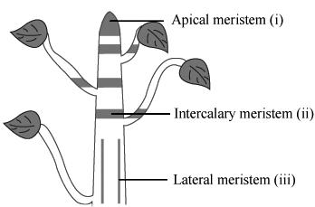
How many types of meristems are present in plants, on the basis of position?
On the basis of location The meristem can be classified into three types:
(i) Apical meristem is present at the tip of stem, root and their branches.
(ii) Intercalary meristem is found at the leaf base, above or below the nodes.
(iii) Lateral meristem is found in vascular bundles and known as cambium. The meristematic tissue, which may appear in most of dicot plants, is known as cork cambium.
Sponsor Area
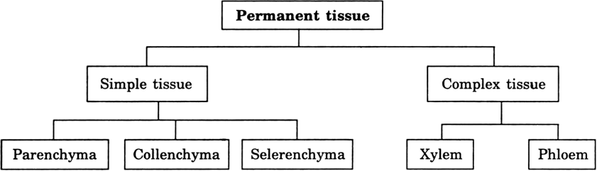
What are plant permanent tissues?
Plant permanent tissues are group of cells which have lost the ability to divide. They are produced by meristematic tissue but lose the ability to divid and are differentiated to perform particular function/functions.
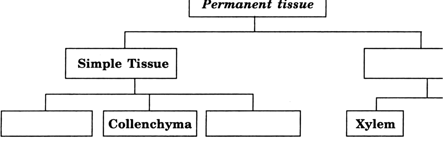
What are simple permanent tissues?
Simple permanent tissues consists of one type of cells only. The cells are similar in origin, structure and functions. For example parenchyma, collenchyma etc.
Name three types of simple permanent tissues.
These are Simple permanent plant tissues:
(i) Parenchyma
(ii) Collenchyma
(iii) Sclerenchyma.
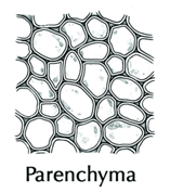
Explain the structure of parenchyma. What are its major types?
Parenchyma. It is the basic or fundamental tissue found in plants. Parenchyma acts as a packing tissue and therefore, it is found in between other tissues. Parenchyma tissue cells store starch, proteins, minerals etc.
i. These are thin walled, circular or polygonal and isodiametric, i.e., having similar sides.
ii. They are living and consists of cytoplasm and a nucleus.
iii. There are many small or a single big central vacuole.
iv. They may not have intercellular space or may have intercellular spaces.
Some parenchymatous cells have pigmented chromoplats. The most important are the cells which contain a number of chloroplasts and the tissue is green, it is known as chlorenchyma.
Some non-green chromoplasts provide different colours to flowers and fruits.
Sometimes the cells contain big air spaces in between them. Such tissue is known as aerenchyma. On the other hand some tissues are green, containing many air spaces then it is called spongy tissue.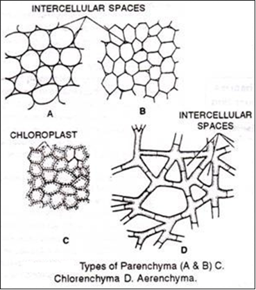
Write the main functions of parenchyma.
The arenchyma performs the following functions.
(i) They store and assimilate food.
(ii) They provide mechanical support as they maintain the turgidity of cells.
(iii) Store waste products such as tannin, gum, crystals of chemicals etc.
(iv) Chlorenchyma containing chloroplasts prepare food through photosynthesis.
v) Parenchymatous cells containing non-green chromoplasts contain pigment that give characteristic colour to flowers (e.g., petals) and fruits.
What are collenchyma cells? Explain their structure with the help of labelled diagram. What is its major function.
Collenchyma ia a type of simple permanent tissue The cells in this type of tissue are living, elongated having irregularly thickened corners. The thickening of the walls are due to cellulose and pectin. They have very little intercellular spaces. These tissues provide flexibility and mechanical support to the plant.
What are the main functions of collenchyma?
The main functions of chollenchyma are to provide mechanical support, tensile strength and elasticity flexibility to the organ in which they are present.
Describe the structure of sclerenchyma. Write its major functions.
Sclerenchyma. Sclerenchyma (scleras-hard) is the chief mechanical tissue of plants. It is a permanent tissue.
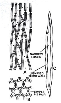
Structure : The cells are made up of sclerenchymatous tissue. The cells are usually long, narrow, pointed at both ends and uniformly thickened by the deposition of lignin without any space in between the cells. The walls are often very highly thickened so that the lumen or cell cavity is nearly obliterated. They are usually provided with simple pits which may be oblique or straight.
Characteristics : The cells of the sclerenchyma are dead. The tissue makes the plant hard and stiff. The sclerenchyma cells are also known as fibres or sclerenchyma fibres, since they are elongated thread-like.
Location : The tissue is present in stems and vascular bundles, in veins of leaves and in the hard covering of seeds and nuts.
Function : Sclerenchyma fibres give necessary strength, rigidity and flexibility to the plant body. Fibres are thick walled and excellent textile fibres of commercial importance such as jute, sunn hemp.
Is the outer most protective layer is continuous? If not, what interruptions are usually found on leaves and herbaceous parts of the plant?
The outermost layer of cells i.e., usually the epidermis is not continuous at some places e.g., on the surface of leaves or green herbaceous stems.
The epidermis of the young shoot and leaves contains numerous minute pores called stomata. Each stomatal opening is surrounded by two semilunar cells known as the guard cells.
The term ‘stoma’ (plural-stomata) is applied to the stomatal opening plus the guard cells. The guard cells are living and contain chloroplasts. Their inner walls (wall towards opening) are thicker and outer walls thinner. The guard cells regulate the opening and closing of the stomatal pore.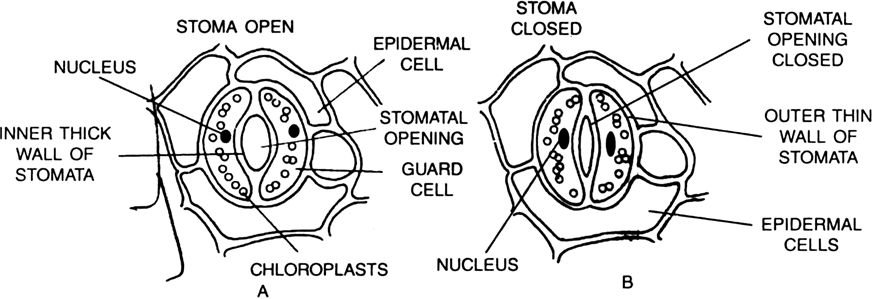
Write major functions of stomata present in the epidermis.
Major Functions of Stomata are:
(i) Stomata are essential for exchange of gases between the plant and the atmosphere.
(ii) Normally, plants eleminate excessive water in the form of vapour through stomatal openings. The process is called Transpiration.
(iii) When there is shortage of water, stomatal openings get closed to reduce water loss. Stomatal openings also close down during night. Thus, stomata regulate water loss from plants i.e., they regulate transpiration.
What is the difference between:
(1) Meristematic cells and permanent cells.
(2) Parenchyma and collenchyma.
(1) Difference between Meristematic cells and Permanent cells.
|
Meristematic Cells |
Permanent Cells |
|
1. They have dense cytoplasm and a large centrally placed nucleus. |
1. They have less amount of cytoplasm, large central vacuole, and normal nucleus. |
|
2. These cells are capable of dividing new cells. |
2. They attain permanent shape and are not capable to produce new cells. |
(2) Difference between Parenchyma and Collenchyma.
|
Parenchyma |
Collenchyma. |
|
1. It is a living tissue. Its cells store may store waste products such as tanin, resins, crystals etc. |
1. It is livingtissue and provides tensile strength the organ in which they are present. |
|
2. Parenchymatous cells may or may not have intercellular shape. Their walls do not have thickening at the corners. |
2. Collenchymatous cells, usually do not have intercellular spaces. They have thickening at the corners of cell walls due to deposition of cellulose and pectin. |
Differentiate between collenchyma and sclerenchyma.
These are difference between collenchyma and sclerenchyma.
|
Collenchyma |
Sclerenchyma |
|
1. The cells of collenchyma are living and have the cytoplasm and the nucleus. |
1. The cells are dead. They do not have the cytoplasm and the nucleus. |
|
2. The collenchyma cells have thickening of cellulose and pectin at the corner of its cells. |
2. The sclerenchymatous cells do not have such thickenings. |
|
3. They provide mechanical support and elasticity to the plant organ. |
3. They mainly provide mechanical support to plant and rigidity to the plant. |
|
4. Collenchyma cells may contain chlorophyll and can also help in the manufacture of starch and sugar. |
4. They do not contain chlorophyll in any condition as they are dead cells. |
What is the difference between sclerenchyma fibres and sclereiods?
Difference between sclerenchyma fibres and sclereiods.
|
Sclerenchyma Fibres |
Sclerenchyma Sclereiods |
|
1. They are dead sclerechmatous cells which are long, pointed at both end and have lignified cells. |
1. They are dead sclerenchymatous cells which do not have definite shape. |
|
2. Their main function is to give mechanical support. |
2. They provide local mechanical support and rigidity to the part where they are present. |
What are complex plant tissues?
A complex tissue is made up of more than one type of cells which work together in coordination as a unit. For example - xylem and phloem .
What is the main function of vascular tissues in plants?
Vascular tissues in plants perform the function of transport. The xylem tissue transports—(i) water and dissolved minerals from roots to various parts plant body. While the phloem transports prepared food material from leaves to different plant parts.
What are vascular bundles?
The conducting tissue like xylem and phloem together form the vascular bundles.
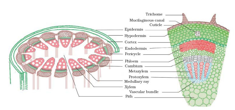
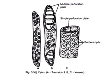
What is xylem? Explain its structure. Which one of its component is very important and why?
Xylem is a complex plant tissue or vascular tissue. Xylem consists of four elements that are-:
(i) Tracheids. A tracheid is an elogated hollow cell with its both ends tapering. The walls of these cells are thick by the deposition of lignin. At certain spots lignin is not present.These spots are termed as pits. These cells are arranged in such a fashion so as to form a system of long tubes and channels in which water can move readily. The tracheids are dead cells. The walls show various kinds of thickenings.
(ii) Vessels are tube like structures formed by a number of cells placed end to end with their transverse walls dissolved or absorbed. The side walls of these tubes also have deposition of lignin. The thickening of the walls show various kinds of patterns. They are also dead cells.
(iii) Wood (xylem) Parenchyma. These are parenchymatous, thin walled living cells. They help in short term conduction of materials. They also store starch and fatty materials.
(iv) Wood Fibres. Xylem sclerenchyma consists of lignified dead fibres. They provide mechanical support.
The most important element of xylem is vessel because they conduct most part of water and mineral salts upward from roots to different parts of shoots.

Fig. 6.10. Tracheids of xylem.

List four component elements of phloem. Which one of them is most important and why?
Phloem consists of the following four elements:
(i) Sieve tubes (ii) Companion cells
(iii) Phloem parenchyma (iv) Phloem sclerenchyma.
The most important element of phloem is sieve-tubes because they translocate food materials from leaves to other parts of plant.
Describe the structure of phloem.
Structure of Phloem. Phloem is a complex permanent tissue of the plant. The phloem consists of four types of elements: sieve tubes, companion cells, phloem fibres and the phloem parenchyma
The main conducting part of the phloem is sieve tube which is formed of elongated cylindrical cells arranged in vertical rows. The terminal walls of each sieve tube member has many minute pores through which food material passes very easily. The entire porous plate is termed as sieve plate. Each sieve tube member is supported by a long parenchymatous cell called the companion cell which helps the sieve tubes in the conduction of food material.
Phloem parenchyma formed of parenchymatous cells. They store the food material. Phloem also contains phloem fibres, also known as Bast Fibres. 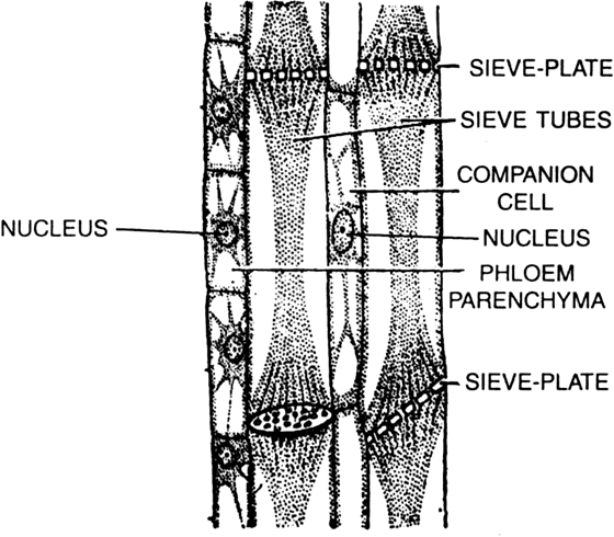
A longitudinal section of phloem.
Differentiate between:
(i) Xylem and phloem.
(ii) Vessel and sieve tube.
(iii) Tracheid and vessel.
(i) Xylem and Phloem.
|
Xylem |
Phloem |
|
1. It consists mainly the dead tissue (except xylem parenchyma). |
1. It mostly has living tissue (except phloem fibre). |
|
2. It is composed of tracheids, vessels, xylem parenchyma and xylem fibre, (in stems) |
2. It is composed of sieve tube, sieve plates, companion cell, phloem parenchyma and phloem fibre. |
|
3. It transports water and mineral from roots to other parts of the plant. |
3. It transports prepared food from leaves to the other plant parts. |
|
4. The flow is unidirectional. |
4. Flow is bi-directional. |
(ii) Vessel and Sieve Tube.
|
Vessel |
Sieve Tube |
|
1. It is a part of Xylem tissue |
1 It is a part of phloem tissue. |
|
2. It is a long distance channel made up of a number of dead cells with hollow lumen. |
2. It is a long distance channel made up of a number of living cells with vacuolated cytoplasm. |
|
3. Vessel conducts water and mineral salts. |
3. Sieve tube conducts food or organic nutrients. |
|
3. The wall is lignified. |
3. The wall is unlignified. |
|
4. The wall is thick. |
4. The wall is thin. |
|
5. The end walls are commonly dissolved completely. |
5. The end walls are perforated to form sieves plate. |
|
6. Besides conduction of sap, a vessel provides mechanical strength. |
6. It does not provide any mechanical strength. |
(iii) Vessel and Tracheid.
|
Vessel |
Tracheid |
|
1. A vessel consists of a large number of cells fused together in a single file. |
1. A tracheid consists of a single cell. |
|
2. The ends are rounded. |
2. The ends are pointed. |
|
3. The septa between adjacent cells of a vessel are usually absent. |
3. The walls between adjacent tracheids, remain intact. |
|
4. The vessel is quite long (1-6 metres). |
4. The tracheid is comparatively short (generally 1 mm). |
|
5. The wall is less thickened. |
5. The wall is more thickened. |
|
6. The lumen is wide. |
6. The lumen is narrow. |
In some plants like desert plants, epidermis has a waxy substance of cutin on its outer surface. Can you think why?
Cutin is a waxy substance that has waterproof quality. It prevents loss of water and helps check loss water during transpiration in desert plants.
Name types of simple tissues.
The three types of simple permanent tissues are
(i) Parenchyma
(ii) Collenchyma and
(iii) Sclerenchyma.
What are the constituents of phloem?
Phloem is made up of four types of elements : Sieve tubes, companion cells, phloem fibres and the phloem parenchyma.
What are the four important type of tissues found in higher animals including man?
The four animal tissues found in higher animals including man are:
(i) Epithelial tissues
(ii) Connective tissues
(iii) Muscular tissues
(iv) Nervous tissues.
What are epithelial tissues?
Epithelial Tissues is the tissue that forms a protective covering on an external or internal surfaces of the animals. The cells are tightly packed and form a continuos sheet. These tissue have no intercellular spaces and have small amount od cementing material between them. An epithelial tissue may be composed of one (simple epithelium) or more layers of cells (compound epithelium). The skin, surface layers of mouth, alimentary canal, lungs etc. are made of epithelial tissues. It also forms a barrier to keep different body system separate and also regulates the exchange of materials between the body and the external enviroment.
What are the five main functions of simple epithelial tissues?
The main functions of simple epithelial tissues are:
(i) The cells form the outer layer of skin. They protect the underlying cells from drying, injury, infection and chemical effects.
(ii) They regulate the exchange of materials between the body and the external environment.
(iii) They help in absorption of water and other nutrients, especially in alimentary canal.
(iv) They help in the elimination of waste products.
(v) Some of them are greatly specialized (glandular cells) and perform secretory function.
What are the categories of epithelial tissue depending upon the shape and function of epithelial cells.
The epithelial cells depending upon the shape and function of the cells are classified into 3 types:
i. Squamous epithelium.
ii Cuboidal epithelium.
iii Columnar epithelium.
What are the various forms of cells of epithelial tissue? Describe briefly.
Following are the different types of epithelial tissue:
(i) Squamous epithelium. The cells of this tissue are broad, flat forming a pavement like appearance. They are of two types Simple squamous( having a single layer) and stratified squamous ( having many layers). It is found in the alveoli of the lungs. Bowmans capsule, membranous labyrinth of internal ear, peritoneum of coelom and blood vessels, skin etc.
(ii) Cuboidal epithelium. The cells of this tissue are cube like in appearance. They usually serve as gland cells. Cuboidal epithelium is called glandular epithelium when a portion of the cuboidal tissue moves inward and forms a multicellular gland. It is found in lining of kidney tubules and salivary glands etc.
(iii) Columnar epithelium. The cells of this tissue are more tall than wide, placed side by side. Their nuclei are situated near the bases. They may have on their free surfaces finger like projections—the microvilli. This tissue usually lines the internal surfaces of stomach and intestine.
Ciliated columnar epithelium. The cells of this tissue are modifications of columnar epithelial cells. They have on their free ends many small, vibratile cilia. These are found in kidney tubules, trachea oviduct etc.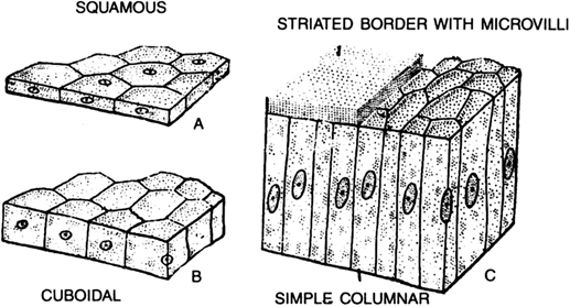
A. Squamous. B. Cuboidal. C. Columnar.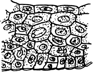
Striated squamous epithelium.
What are muscular tissue. What is their function?
Muscular Tissue are composed of contractile, fibre-like cells. Intercellular substance is very little. The cells of muscular tissue remain enclosed or surrounded by connective tissue.
Functions. The mescle tissue is responsible for the movement in out bodies. The movement of the body or limbs is brought about by contraction and relaxation of muscle cells.
What are the three types of muscle fibres (muscle cells)?
There are three types of muscle fibres:
(i) Striated muscle (skeletal muscle or voluntary muscle).
(ii) Unstriated muscle (smooth muscle or involuntary muscle).
(iii) Cardiac muscle.
Explain the structure of three types of muscle fibres. Also write the locations where they are found in the body.
The following are the three types of muscle cells:
(i) Smooth muscle fibres :They are also known as Unstriped muscles. They are involuntary in nature. This type of muscular tissue consists of spindle-shaped, long fibre-like cell with a nucleus at the centre. The ends are undivided. This type of muscles is present in alimentary canal, muscular coat of blood vessels etc.
(ii) Striated muscles. (Also known as striped or voluntary muscles because of their function being in our control or will). This type of muscular tissue cells are very long fibres, enclosed in a membrane known as sarcolemma. The cells (fibre) are multi-nucleated. Each fibre has several longitudinal myofibrils embedded in cytoplasm. On these myofibrils are present characteristic light and dark bands which give it a striated appearance. These muscles are attached to the skeleton; so are also called skeletal muscles,
(iii) Cardiac muscles. These muscles are found in heart. They are not under the control of the will. They contract rhythmically and involuntarily throughout life without the sign of fatigue.
Structurally they show the characters of both unstriated and striated muscles. They are made up of branched fibres. These fibres are multinucleated and show alternate light and dark bands (striation).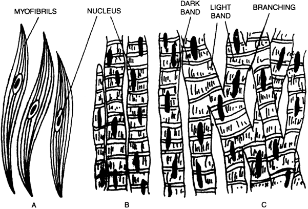
Muscular tissue —A. Unstriped. B. Striped. C. Cardiac.
What is basement membrane?
Basement membrane is a very thin non-cellular fibrous membrane on which epithelial tissues rest. It also separates the tissue from the underlying tissues.
Write structure, location and function of squamous epithelium, cuboidal epithelium, columnar epithelium and striated squamous.
| Type of tissue | Structure | Location in the body |
Function |
|
| 1. | Squamous epithelium | Cells are flat, discoi-dal and fit together forming a pavement like appearance. | Alveoli of the lungs Blood capillaries. Bowman’s capsule of nephron in kidney. |
Exchange of gases in the lungs. Exchange of materials and gases between body cells and blood. Ultrafiltration of blood. |
| 2. | Cuboidal epithelium | Cells are cuboidal with centric and rounded nuclei. |
Tubule of nephron in kidney. Duct of salivary glands. |
Selective reabsorption of useful materials and water. Production of gametes. |
| 3. | Columnar epithelium | Cells are more tall than wide, placed side by side. The nucleus is situated near the bases. |
Fallopian tube (ciliated cells). Mucosa of small intestine (cells with microvilli). |
Help in movement of ovum/zygote. Absorption of digested food with increased surface area. |
| 4. |
Striated squamous |
Squamous flat cells arranged in many layers to prevent wear and tear of parts. |
Skin, tongue, oesophagus lining of mouth. |
Protection, prevent wear and tear. |
Of which type of muscles the stomach wall is made? Whether they are voluntary or involuntary ?
Stomach walls are made up of smooth or non-striated muscles. They are involuntary in nature.
What is the difference between voluntary and involuntary muscles. Give example of each.
Voluntary muscles are those which are directly under our control of our conscious will e.g., skeletal muscle. They work or move on our command.
Involuntary muscles are not directly under command of our will. We cannot stop contraction or restart contraction stomach, intestine or heart muscles. Example. Cardiac and smooth muscles.
Write main characteristic features of skeletal, smooth and cardiac muscles.
| S.No. | Character | Skeletal (Striated) Muscles |
|
Cardiac Muscles | |
|
1. |
Shape of Cells | Cells are long cylindrical, non-tapering and unbranched. | Cells are long with tapering ends (spindle shape) and unbranched. | Cells are non-tape-ring, cylindrical and branched. | |
| 2. | Nucleus | Many nuclei (multinucleated) which are situated towards the periphery of muscle fibre. | The cells have only one nucleus (uni-nucleated) situated in the centre. |
Each cell contains one or two nuclei situated in the centre. |
|
| 3. | Striatation |
|
Stritations or stripes are absent. | Cells have faint stritations. | |
| 4. | Location | Hands, legs and other skeletal muscles. | Wall of stomach, intestine, ureter, bronchi etc. | Present in heart. | |
| 5. | Mode of Contraction | Voluntary (work upon our will) contract rapidly but soon undergo fatigue. | Involuntary not under our will. Contract comparatively slow but do not fatigue. |
|
What is the function of connective tissue?
Connective tissue performs the functions of binding, providing support, transport and connecting together different organs of the body.
What are the two main features of connective tissue?
The two main features of connective tissue are
i. Cells are loosely spaced.
ii. The cells are embedded in the matrix which may be jelly like, fluid, dense or rigid depending upon the function the tissue has to perform.
hat are three main categories of connective tissue?
Categories of Connective Tissue:
(i) Connective Tissue Proper. In this type of tissue there is a matrix in which generally two types (white and yellow) fibres are present. In between these fibres some other cells are present in the matrix.
(ii) Skeletal Tissue. This type of tissue forms the skeleton of an organism. It may consist of notochord, cartilage and bone.
A cartilage has a matrix, called chondrin, in which a network of collagen fibres is present. There are present cells known as chondrocytes. There may be present empty spaces filled with fluid known as lacunae.
In bones, matrix is formed of a protein called ossein impregnated with phosphate and carbonates of calcium and magnesium.
(iii) Fluid Tissue. Blood and lymph tissues are called fluid tissue. These are specialized connective tissue. It consists of liquid matrix with no fibres. In this liquid matrix-plasma there are corpuscles. Blood transports food material, gases and other substances to the various parts of the body of an animal.
List five examples of connective tissue.
Examples of connective tissue : (i) Bone (ii) Carliage (iii) Tendons (iv) Ligaments (v) Areolar (vi) Adipose and (vii) Blood.
Describe the structure of cartilage and bone.
Cartilage : It is a solid but semi-rigid and flexible connective tissue. It has large, bluntly angular cartilage cells called chondriocytes. They occur in clusters of 2 or 3 in small spaces (lacunae) scattered in the matrix. The cartilage is bounded externally by a sheath called perichondrium.
Hyaline cartilage is found in tracheal and broncheal rings, fibro-cartilage is present in intervertebral discs and pubic symphysis, elastic cartilage is found in pinna, tip of nose and eustachin tube.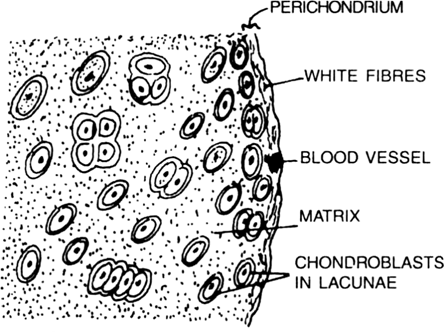
T.S. of Cartilage.
Bone : Bone is solid, rigid and strong connective tissue. Its matrix become hard due to the deposition of salts of calcium and phosphorus. Osteocytes or bone cells are present in irregular spaces—lacunae in the matrix, interconnected by fine canals called canaliculi. The matrix occurs as layers—the lamellae arranged in concentric rings around narrow longitudinal cavities called Haversian canals. These canal carries blood vessels and nerves.
The haversian canals are inter-connected by transverse channels called Volkman’s canals. The long bones have central cavity filled with bone marrow and is called marrow cavity. The bone marrow is formed of blood vessels, nerve fibres and adipose tissue. Blood cells are produced in bone marrow. The bone is bounded externally by a tough sheath called periosteum.
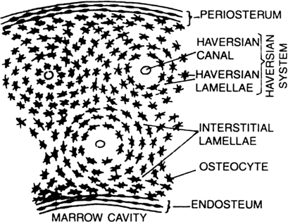
T.S. of bone.
Explain the structure of a fluid connective tissue blood.
Blood is a fluid connective tissue. It consists of (i) Blood plasma, (ii) Blood corpuscles.
(i) Blood plasma. It forms generally fluid matrix which contains 85 to 90% water, 7% different types of proteins, 0.9% of salts, about 0.1% glucose and a very small amount of hormones, wastes, etc. In the plasma fluid blood corpuscles (Cells) are found suspended.
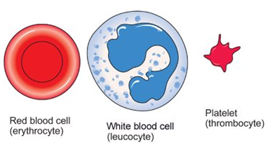
(ii) Blood corpuscles. Three kinds of blood corpuscles are found suspended in the blood plasma. These are : (i) Red blood corpuscles (Erythrocytes) or RBC (ii) White blood corpuscles (Leucocytes) or WBC and (iii) Blood platelets.
(i) Red Blood Corpuscles (Erythrocytes) or RBC
The red blood corpuscles are biconcave disc-like structures devoid of nuclei.
(ii) White Blood Corpuscles (Leucocytes) or WBC
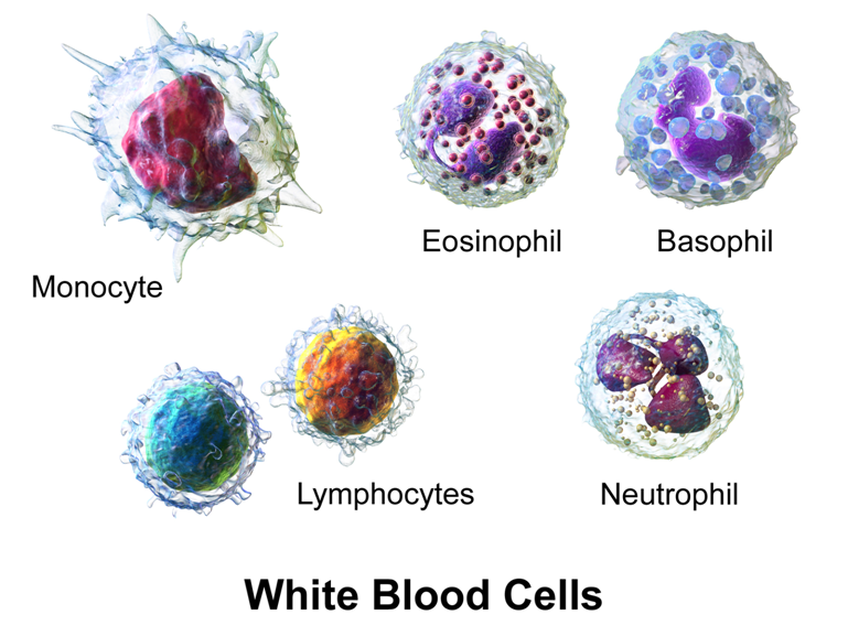
These are comparatively smaller in size, semi-transparent, colourless, irregular in appearance. These are subdivided into following categories: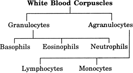
Granulocyte. These are irregular amoeboid leucocytes. Their cytoplasm contains many small granules hence the name granulocytes.
(a) Basophils. These are small. Their nucleus is usually multilobed.They are strained with basic dyes.
(b) Eosinophils. They have a bilobed nucleus. They can be stained with acid dyes.
(c) Neutrophils. Their nucleus is multi-lobed.
Agranulocytes. As the name suggests their cytoplasm does not contain granules. They have a large nucleus and are of two types.
(a) Lymphocytes. Size 7-8, 10-12 μ. They are round, slow moving and phagocytic in functions.
(b) Monocytes. Size 12-20 μ. They have kidney shaped nucleus. They are slow moving and phagocytic.
(iii) Blood Platelets
These are small, 2-4 μ in diameter. They are without nucleus. Their main function is to liberate thromboplastin which causes blood clotting.
What are Areolar tissue and Adipose tissue? Where they are located?
i. Areolar Tissue. It is a connective tissue. It consists of matrix, several types of cell, collagen and elastin fibres.
Location : They are found between the skin and muscles, around blood vessels and nerves and in the bone marrow.
Functions : It fills space inside the organ, support internal organ and helps in repair of the tissues.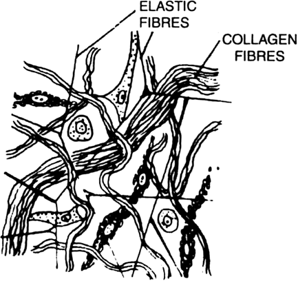
Areolar Tissue.
ii. Adipose Tissue. It consists of cells (adipocytes) which are filled with fat globules, matrix, collagen and elastin fibres.
Location. It is found below the skin and between internal organs.
Function. It stores fats and act as insulator.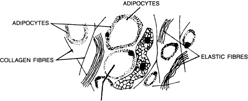
Areolar tissue.
Give two differences between striated and unstriated muscles.
|
Striated Muscles |
Unstriated Muscles |
|
1. These are cylindrical, with non-tapering ends. |
1. These are spindle shaped and have pointed tapering ends. |
|
2. Transverse alternate light and dark bands or striatations can be seen. |
2. No light and dark bands or striatations are seen. |
|
3. There are many nuclei (multinucleated), these nuclei are situated towards the periphery of the muscle fibre. |
3. The cell has only one nucleus (uninucleated) situated in the centre. |
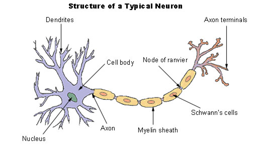
What is nervous tissue?
Nervous Tissue. Nervous tissue is made up of nerve cells or neurons.
Each neuron has a single long part called the axon and many short branches celled the dendrites.
The cell body contains a distinct nucleus and has a special kind of granule, called Nissl’s granule in the cytoplasm.
In some nerve cells the axon has a thick white covering called medullary sheath. These are called medullated fibres. When this sheath is absent, it is called non-medullated fibres.
In the medullated fibres, the sheath is not continuous—there are gaps along the entire length. Each gap is called Nodes of Ranvier.
The terminal end of axon is without sheath and branched. The axon of one neuron remains in contact with the branches of dendrites from another neuron. The region of contact is called synapse.
Each nerve cell receives message through the dendrites and sends message through the axon.
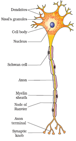
Nerve cell.
How would you recognize that the slide given to you is of striated muscle ? Give only two important points?
Striated muscels have a distinct characteristics
(i) Striated muscles have light and dark bands (which gives it a striped look) (ii) Striated muscle fibres are not branched like cardiac muscles.
Which cell may be the longest in the body of an animal?
Neuron (Nerve cell) is the longest cell which may be 1 m to 2 m long (in special cases).
Why is blood called as fluid connective tissue?
Blood contains liquid matrix and blood cells are floating in it. It does not have fibres. It circulates in the body. Since it forms the framework that support the body. It anchors the muscles and supports the body therefore it is calle a connective tissue.
Why are striated musclels generally called skeletal muscles?
In general, striated muscles are found attached to the bones. They help in the movement of the bones and so, they are also called skeletal muscles.
Differentiate between tendon and ligament.
|
Tendon |
Ligament |
|
1. It attaches bone skeletal muscle to a bone. |
1. It connects a bone with another bone. |
|
2. It contains thick parallel bundles of white collagen fibres. |
2. It contains both white collagen and yellow elastic fibres running in different direction. |
|
3. It is non-elastic . |
3. It is elastic. |
|
4. Less flexibility |
4. More flexibility. |
Give three features of cardiac muscles. Give a simple diagram.
Following are the three features of cardiac muscles:
i) Cardiac muscle cells are cylindrical branched and uninucleate.
(ii) The show rhythmic contraction and relaxation throughout life.
(iii) They are involuntary in nature .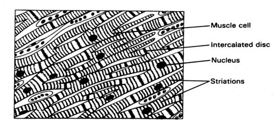
What are the functions of areolar tissue?
Areolar tissue fills space inside the organs, support internal organs and help in repair of the tissues.
Sponsor Area
Sponsor Area