Explain the structure of a fluid connective tissue blood.
Blood is a fluid connective tissue. It consists of (i) Blood plasma, (ii) Blood corpuscles.
(i) Blood plasma. It forms generally fluid matrix which contains 85 to 90% water, 7% different types of proteins, 0.9% of salts, about 0.1% glucose and a very small amount of hormones, wastes, etc. In the plasma fluid blood corpuscles (Cells) are found suspended.
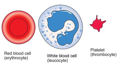
(ii) Blood corpuscles. Three kinds of blood corpuscles are found suspended in the blood plasma. These are : (i) Red blood corpuscles (Erythrocytes) or RBC (ii) White blood corpuscles (Leucocytes) or WBC and (iii) Blood platelets.
(i) Red Blood Corpuscles (Erythrocytes) or RBC
The red blood corpuscles are biconcave disc-like structures devoid of nuclei.
(ii) White Blood Corpuscles (Leucocytes) or WBC
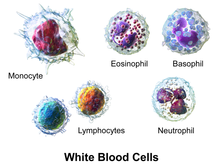
These are comparatively smaller in size, semi-transparent, colourless, irregular in appearance. These are subdivided into following categories: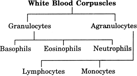
Granulocyte. These are irregular amoeboid leucocytes. Their cytoplasm contains many small granules hence the name granulocytes.
(a) Basophils. These are small. Their nucleus is usually multilobed.They are strained with basic dyes.
(b) Eosinophils. They have a bilobed nucleus. They can be stained with acid dyes.
(c) Neutrophils. Their nucleus is multi-lobed.
Agranulocytes. As the name suggests their cytoplasm does not contain granules. They have a large nucleus and are of two types.
(a) Lymphocytes. Size 7-8, 10-12 μ. They are round, slow moving and phagocytic in functions.
(b) Monocytes. Size 12-20 μ. They have kidney shaped nucleus. They are slow moving and phagocytic.
(iii) Blood Platelets
These are small, 2-4 μ in diameter. They are without nucleus. Their main function is to liberate thromboplastin which causes blood clotting.